Surgical Oncology
Modern cancer treatment requires the Surgical Oncologist to be well versed with a wide range of procedures starting from a small but important procedure like a biopsy to some of the most complex Surgeries ever carried out on the human body. At KIMS we ensure that the most basic as well as most complex, advanced surgical oncology services are tailored to the specific needs and preferences of each patient. Biopsy is done by conventional methods or minimally invasive techniques like Trucut, USG or CT guided, endoscopy etc. Radical Surgical procedures are performed for solid tumors and cancers of most parts of the body from head to toe by conventional methods or by minimally invasive means like laparoscopy, thoracoscopy. We are equipped with world class equipment and supported by in-house speciality and super speciality faculty and facilities like Intensive care, Plastic and reconstructive surgery, Gastroenterology, Advanced and interventional Radiology, Immunohistochemistry etc. that have enabled us to give results comparable to the best in the world over the last 5 years.
Faculty:
Two well trained and experienced Surgical Oncologists have been available at KIMS for the past 5 years. Here, they have successfully performed most of the procedures required for the management of solid tumors, cancers of the human body .This includes some of the most complex Surgeries ever carried out by surgeons worldwide .Earlier, such procedures required people from our region to travel to centres outside for treatment . All this has been achieved with extremely low morbidity, mortality and recurrence rates. With a combined experience in oncology of about 17 years and an impeccable track record, they are probably the best team to help you out.
Dr. Saroj Ranjan Sahoo, received both his M.B.B.S. and M.S. Degree in Surgery from Utkal University. He has worked as a senior resident in Cancer Surgery at Safdarjang Hospital, New Delhi before moving on to Tata Memorial Centre, Mumbai. There he received his training as a Senior Resident in Surgical Oncology for 3 years under the leading Surgical Oncologistsof the country. He joined Asian Hospital, Faridabad as a Consultant Oncosurgeon where his skills and services were highly appreciated. With an intention to return to his roots, he joined KIMS in 2014 and has been working as a Consultant, Surgical Oncology at Pradyumna Bal Memorial Hospital, Kalinga Institute of Medical Sciences.
Dr. Sabyasachi Parida, is a Gold Medal awardee in M.S. Surgery from Utkal University. He has worked with reputed scholar surgeons of the country at Maulana Azad Medical College, New Delhi after receiving his post graduate degree. With a keen interest in Surgical Oncology he joined the Department of Cancer Surgery at Safdarjang Hospital, New Delhi as a Senior Resident. There he received his training in Oncosurgery for 3 years under one of the most senior, skilled and learned Cancer Surgeons of our country. Upon completion of his training, he moved back to Bhubaneswar in 2013 and has been working as a Consultant, Surgical Oncology at Pradyumna Bal Memorial Hospital, Kalinga Institute of Medical Sciences.
Equipment:
Well equipped with some of the best equipment available worldwide for performing basic and complex surgeries, it also possesses equipment for patient care in prolonged Surgeries. This includes Electrosurgical equipment, ultrasonic scalpel, vessel sealing devices, surgical headlight, table mounted retractor, advanced laparoscopic instruments and equipments, fluid channel warmers, warmer blanket, DVT pump etc. Other than these, allied super speciality departments are well equipped with advanced equipment like operating microscope, heart lung machine, ECMO etc.
Procedures:
Indicative list of procedures available is listed below. New and innovative procedures are regularly being added as per case requirement.
- Head and Neck Surgery: Oral Cavity Cancer and precancerous conditions, Tongue Cancer , Thyroid Cancer and Non-Cancerous lesions, Parotid cancer and Tumors, Submandibular Gland Cancer and Tumors, Parathyroid gland tumors, Laryngeal Cancer etc.
- Upper Gastrointestinal Surgery: Gastric Cancer and Tumors, Duodenal Cancer and Tumors, Jejunal Cancer and Tumors, Ileal Cancer and Tumors, Splenic Tumors.
- Lower Gastrointestinal Surgery: Colon Cancer and Tumors, Rectal Cancer and Tumors.
- Hepatobiliary Surgery: Hepatic Cancer and Tumors, Biliary Cancer and Tumors, Bile Duct Cancer and Tumors.
- Pancreatic Surgery: Pancreatic Cancer and Tumors.
- Retroperitoneal Surgery: Retroperitoneal Sarcomas, Tumors.
- Endocrine Surgery: Adrenal Cancer and Tumors, Ovarian Cancer and Tumors, Thyroid and Parathyroid Tumors.
- Gynaecological Surgery: Ovarian Cancer and Tumors, Uterine Cancer and Tumors, Cancer of Uterine Cervix, Fallopian Tube and Peritoneal Cancer.
- Urogenital Surgery: Kidney Cancer and Tumors, Ureteric Cancer, Penile Cancer, Testicular Cancer and Tumors.
- Transthoracic Surgery: Oesophageal Cancer and Tumors, Lung Cancer.
- Skin and Soft tissue Surgery: Skin Cancer and Tumors,Soft Tissue Sarcoma and Tumors.
- Extremity Surgery: Soft Tissue Sarcoma and Tumors, Bone Sarcoma and Tumors.
- Breast Surgery: Breast Cancer and Tumors.
Publications:
A few interesting, challenging cases which have been published are detailed below:
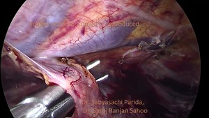
Minimally Invasive Surgery for Cancer of the Esophagus:
The oesophagus( food pipe) is a long muscular tube that constitutes the primary part of the alimentary tract between the oral cavity and stomach. It passes through three body compartments the neck, the chest and the abdomen where it continues into the stomach. In the chest it lies anterior to the vertebral column posterior to the wind pipe, adjacent to major vessels going to and coming from the heart. It is flanked on either side by the lungs and the heart lies to the left. It is for this reason that even the most skilled surgeons have found it difficult to perform surgery for esophageal cancer. Among different parts of the digestive tract, surgery for the oesophagus took the longest time to be developed. Even today, it is a niche science and art performed by only a few centres across the world. Conventional esophageal cancer surgery as we do it today came into being in 1976(Mc Keown) whereas conventional pancreatic cancer surgery had been in vogue since 1940(Whipple).Even today, the number of centres performing esophageal surgery is far fewer than those that perform pancreatic surgery. The major hurdle for access to the oesophagus was the need to open the chest. Even when successfully performed, opening up the chest meant major morbidity for the patient and few were actually able to tolerate it. For those who were able to undergo the procedure, the pain of a chest wound made it difficult for the patient to breathe comfortably during the postoperative period. This meant a heavy dose of analgesics, time in the intensive care unit, longer hospital stay and longer time for rehabilitation. Sometimes, this led to delays in starting adjuvant treatment for esophageal cancer, like Chemotherapy. The development of Minimally Invasive Esophagectomy(MIE) has addressed many of the above issues. De Paula et. al. in 1995 performed the first minimally invasive esophagectomy by transhiatal (through the abdominal) route.Subsequently, transthoracic(through the chest) minimally invasive esophagectomy was developed. When properly planned and executed,it becomes a more tolerable and less morbid procedure for the patient. It is commonly called as video assisted thoracoscopic surgery (VATS) for the oesophagus and is performed well both by the use of laparoscopic instruments as well as by help of surgical slave robot. 4-5 small holes about a cm or less are made in the chest for the purpose. For cancer affecting the middle and lower third of the esophagus, removal of whole of the esophagus with the draining lymph nodes is commonly performed. This involves carefully dissecting it free from its attachments without injuring the vital organs all around it.Dissection in the neck is done to divide and release the esophagus into the chest.Lymp node dissection in the neck might also be required. Careful execution of this part is also critical to the success of the procedure as, even the slightest error can injure nerves to the voice box, severally impairing voice quality. Once the above steps are successfully carried out, the food pipe is retrieved through an upper abdominal incision and replaced by a tube formed from the stomach or rarely, the large intestine. This part of the surgery can be done laparoscopically or by conventional laparotomy (opening the abdomen).If necessary, upper abdominal lymph node dissection is also carried out. The tube so formed is taken up to the neck and sutured with the proximal remaining part of the esophagus. When only the distal part of the esophagus is excised, this anastomosis is carried out inside the chest. As is evident from the above description, it requires a high level of expertise and care to carry out the procedure successfully.
Case Report:
A 60yr gentleman presented to the oncosurgery unit with complaints of difficulty in swallowing food for past 15 days. He was under treatment for hypertension. He was still active professionally and quality of voice was important to the ability to continue in his profession.He had lost weight significantly and hence was thin with an unsatisfactory performance status. He had been evaluated outside with upper G.I. endoscopy whioch had revealed a stricturous growth in lower 1/3rd of his food pipe. The scope was not negotiable further. Biopsy was inconclusive. CECT scan of abdomen and chest had been done which was reported as irregular, circumferential wall thickening involving lower 1/3rd of esophagus reaching upto gastroesophageal (GE) junction, few enlarged perigastric lymph nodes near GE junction. .Repeat Upper gastrointestinal endoscopy was done by our Gastroenterologist and biopsy was done.Disease appeared to be confined to the oesophagus without significant lymph node involvement or distant spread. He also had outside reports that were suggestive of harbouring a contagious hepatitis virus infection. Gastroenterology consultation was sought and further tests were advised which did not reveal any further positive results for infection by a hepatotropic virus. This was an important concern because, if positive, it carried a significant risk of transmission to the surgeon and the surgical team in case of any breach in universal precautions. His frail status and less than satisfactory performance status meant that the proposed surgery carried high risk for him. Further, requirement of preservation of voice quality (tone and timbre) meant that the surgery had to be performed to the highest standards. For such a patient, opening up the chest would have been a too morbid procedure. Hence, it was decided to proceed with minimally invasive esophagectomy through the chest and the abdominal part performed through an upper abdominal incision. This abdominal incision would facilitate retrieval of the resected organ as well as formation of the gastric tube that would replace the esophagus. In view of his frail frame and inability to accept adequate diet, it was decided to place a nasogastric feeding tube and devote a few days to improve his general condition and lung function. This would subsequently enable him to tolerate the procedure better. He underwent the surgical procedure successfully with all the technical parameters achieved. The Surgical team comprised of Dr. Saroj Ranjan Sahoo and Dr. Sabyasachi Parida (Consultants, Surgical Oncology) and ably assisted by a Team of Anaesthetists, OT Technicians, OT Nurses and support staff. The anaesthesia team used a double lumen endotracheal tube to intubate the patient for anaesthesia. This enables selective deflation of one lung (in this case right side) to provide adequate surgical field when required. The chest part was performed with the patient on his left side and body tilted forward. Ports for minimally invasive surgery were placed through the right chest. Esophagectomy and regional lymphadenectomy was done by thoracoscopy. Once the chest part was completed, the patient was turned over to lie on his back for the neck and abdominal part of the surgery. Special care was taken to prevent any possibility of contact of patients body fluids with the surgical team or to other patients (in view of a seropositive report) .Such a major and complicated procedure is seldom performed in patients who are found to be seropositive for a contagious, chronic, incurable viral infection .Only a few centres across the world take up such cases on a routine basis. Although the confirmatory tests were negative in our case, we still took all precautions as part of good surgical practice and carried out the entire procedure without spillage of blood or fluids beyond the operative field. We had the requisite protective gear on us all the time. Waste management was also done with all universal precautions and as per guidelines assuming seropositivity for infection. Immediate postoperative care was effectively provided by the team of Intensivists and Intensive care nurses. Subsequently, competent nurses in the special cabins nursed him back to good health. Laboratory services chipped in with timely and quality reports. The patient recovered relatively quickly considering his unsatisfactory general condition at the beginning. His voice quality is as good as it was preoperatively. He has happily resumed his professional life. He is continuing with further management as per his disease stage. The above example illustrates how careful treatment planning and the highest levels of technical expertise combined with quality support and teamwork can provide the best results even in demanding situations. It should also be mentioned that, not all cases are suitable for minimally invasive esophagectomy and some might require opening up the chest for better control of the surgical field, better disease clearance aided by tactile feedback from the surgeon's fingers and when it is not possible to complete the surgery by minimally invasive means. In some cases preoperative treatment with Chemotherapy, Radiotherapy might be required. All of this is just as important as the surgical procedure itself. Good decision making lies at the heart of every successful outcome in Surgical Oncology.